
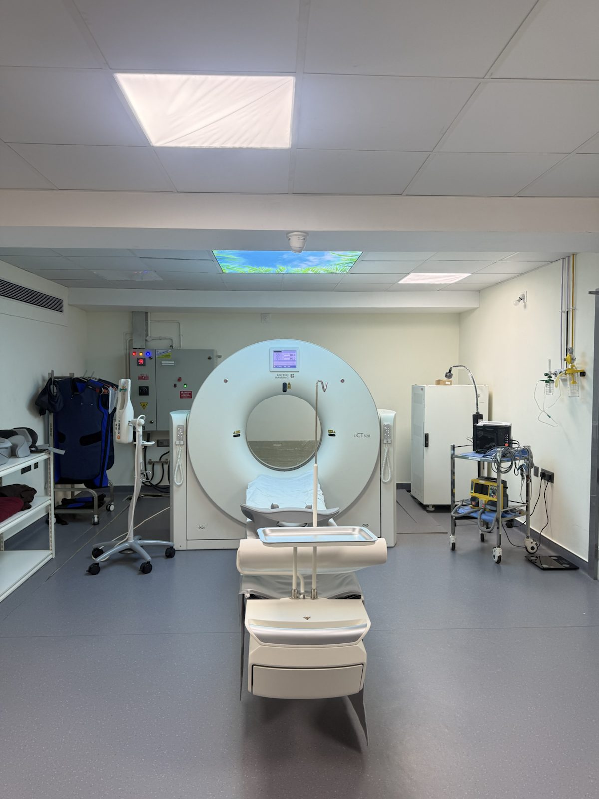
Radiology & Imaging
Digital 1.5T MRI, Digital X-ray, ultrasonography and multi-detector CT scanning supporting outpatient, inpatient and emergency care.
Radiology & Imaging at Mangala Hospital
Radiology supports diagnosis, treatment planning and emergency care across the hospital. Digital 1.5T MRI, Digital X-ray, ultrasonography and multi-detector CT are coordinated in one department, with on-site reporting by a qualified radiologist.
Advanced MRI and CT reconstruction tools support detailed routine and specialised studies, helping clinical teams make timely, informed decisions.
What We Offer
Round-the-Clock Reporting
Imaging support is coordinated with Casualty and inpatient teams for urgent clinical requirements.
View Imaging Facilities →Related Specialities
Need Urgent Care?
Our Casualty & Emergency department is open 24x7, with mobile ICU ambulance backup.
Call: 0824 244 3088Imaging That Keeps Pace With Every Department
Comprehensive Imaging
MRI, X-ray, ultrasound and CT support diagnostic needs across the hospital's clinical departments.
Digital 1.5T MRI
Detailed, radiation-free imaging with dedicated coils and advanced reconstruction technology.
Reconstruction for Clinical Planning
Selected CT studies can be reconstructed to support clinical review and surgical planning.
Reported On-Site
Studies are reviewed by a qualified radiologist so findings can be coordinated with the referring clinical team.
Coordinated Imaging
Imaging and clinical teams coordinate results and next steps within the hospital.
Comfortable, Accessible Care
A convenient, patient-friendly imaging suite designed to make scans as quick and stress-free as possible.
Our Imaging Modalities
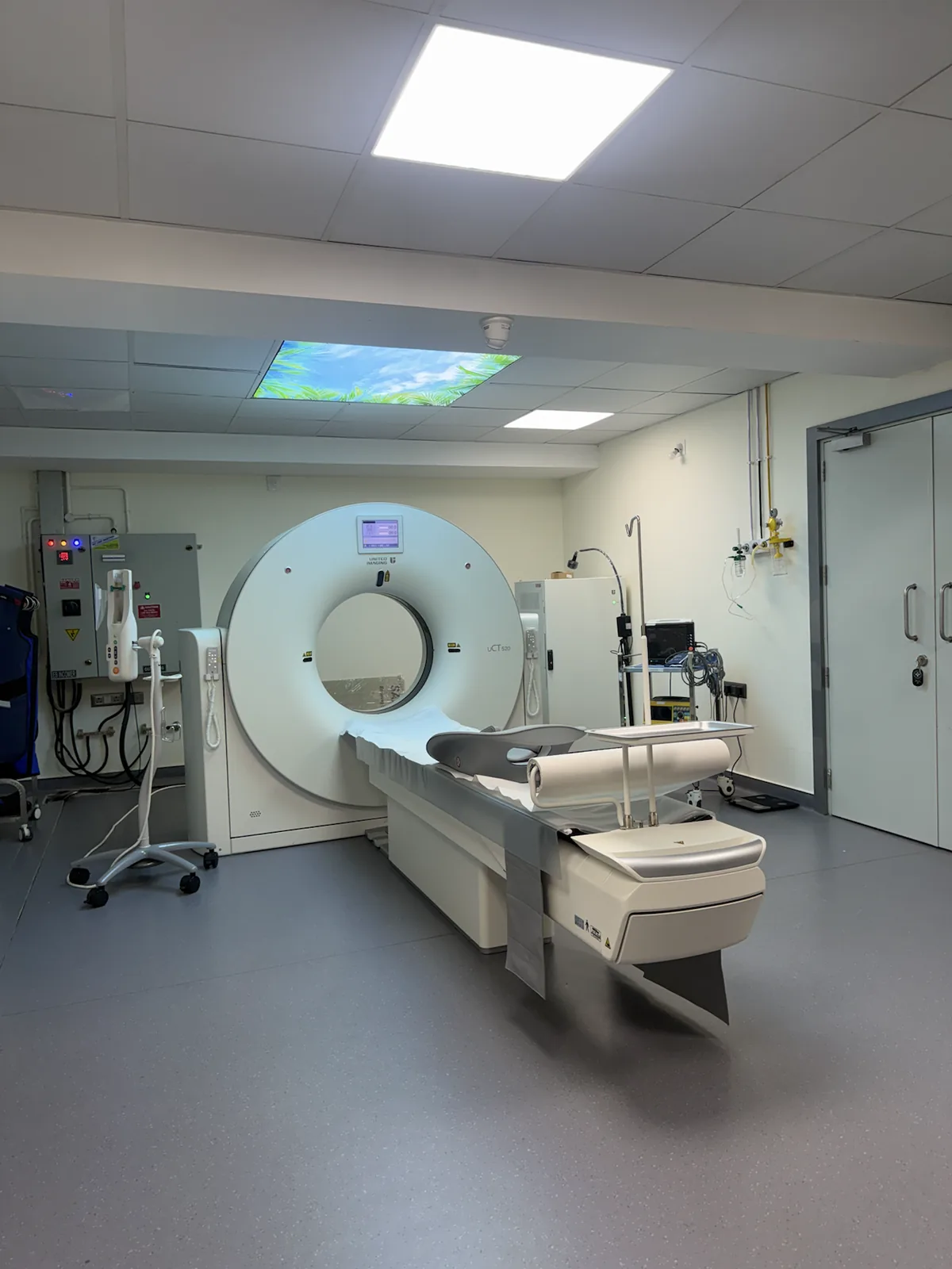
Multi-Detector CT Imaging
Our CT scanner produces cross-sectional images across the brain, chest, abdomen, spine and pelvis, with protocols selected according to the referring doctor's clinical question. Selected studies can also be reconstructed to help clinical and surgical teams review complex anatomy.
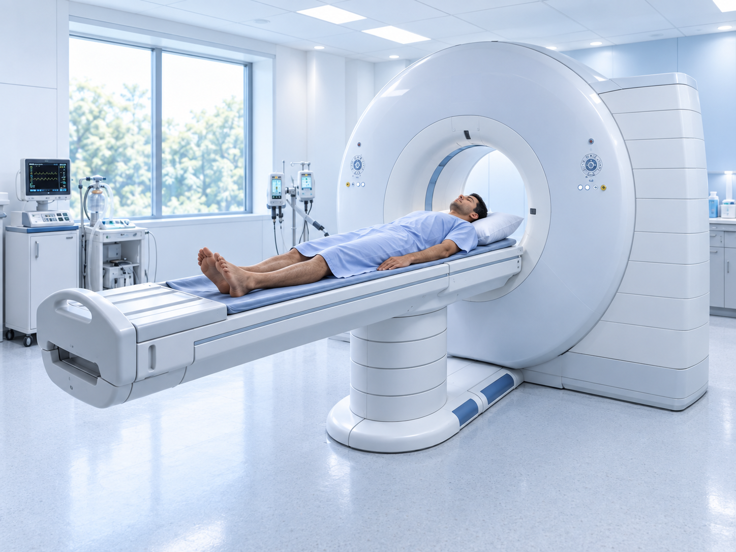
Detailed Imaging Without Radiation
Our Digital 1.5T MRI supports detailed imaging with TIM technology, 16 independent RF channels and dedicated breast and knee coils. It also supports breast MRI with 3D reconstruction for accurate clinical assessment.
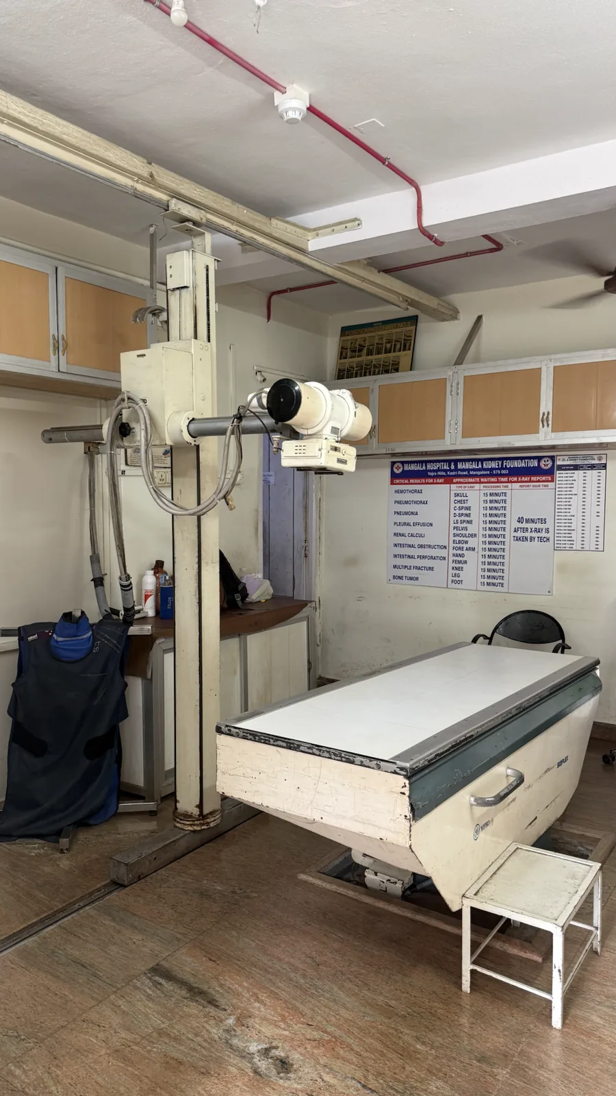
Fast, Clear Imaging for Bones & Chest
Digital X-ray is often the first imaging step for suspected fractures, chest infections, joint injuries and a wide range of everyday diagnostic needs. Prompt image availability is especially useful in Casualty and Orthopaedics, where findings guide the next treatment decision.
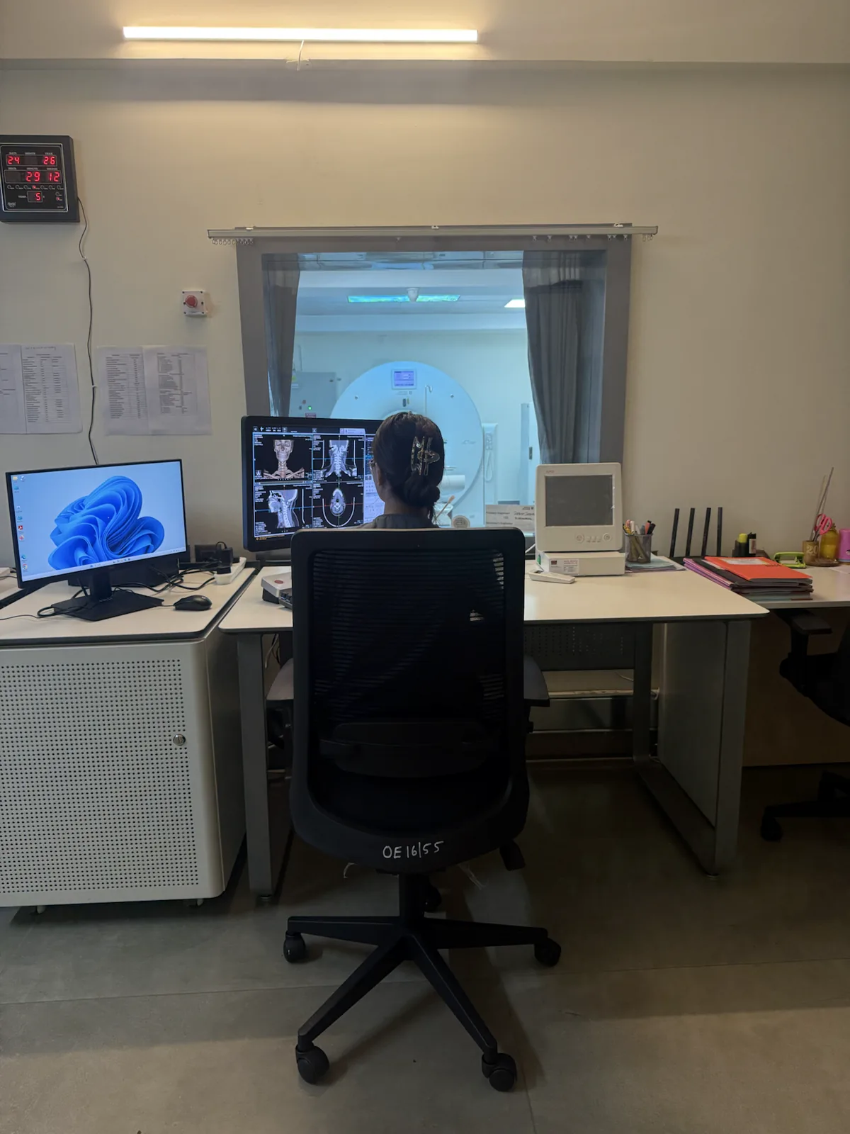
Every Scan, Read Carefully
Every MRI, CT, X-ray and ultrasound study is reviewed on high-resolution reporting workstations before a result reaches the referring doctor. Ultrasonography (USG) is used daily for abdominal and pelvic assessment, soft-tissue evaluation and pregnancy scans — a radiation-free option that works alongside MRI, CT and X-ray. This careful review process keeps reporting timely without sacrificing accuracy.
Meet the Radiologist Leading This Department
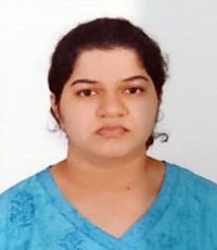
Dr. Swathi P
A radiologist with training in diagnostic imaging, supporting the review and reporting of MRI, X-ray, ultrasound and CT studies across Mangala Hospital's clinical departments.
Our radiology team is supported by trained imaging technologists across MRI, CT, X-ray and ultrasound services.
Your Imaging Journey, Step by Step
Referral or Walk-In
Your doctor requests the scan, or you can walk in directly for select X-ray and ultrasound tests.
Preparation
Some CT and ultrasound studies need fasting or a full bladder — our staff will guide you beforehand.
The Scan
Most X-rays take minutes; ultrasound and CT studies typically take 15-30 minutes depending on the region scanned.
Report & Follow-Up
Studies are reported on-site, with results shared with your treating doctor to plan the next step in your care.
Facilities Supporting This Department
Frequently Asked Questions
Most scans are done on a doctor's referral so the right study and protocol are chosen for your symptoms. Some routine X-rays and ultrasound scans can be done as a direct walk-in — ask our imaging desk to confirm.
CT uses ionising radiation, so the scan protocol is selected according to the body region and clinical requirement. Your doctor and radiology team consider the expected diagnostic benefit before the examination is performed.
Some CT studies (like CECT) and abdominal ultrasound need a period of fasting, and pelvic ultrasound may need a full bladder. Our staff will give you specific instructions when your scan is booked.
Yes. Ultrasound uses sound waves rather than radiation and is the standard, safe imaging method used throughout pregnancy for monitoring the baby's growth and wellbeing.
Reporting time depends on the examination and clinical priority. Emergency and inpatient studies are coordinated with the treating team, and staff will advise when the report is expected.
Yes, diagnostic imaging is covered under most cashless and reimbursement mediclaim plans as part of your overall treatment. See our Insurance page for the full list, or check with our billing desk.